Development of a Virtual AI Testbed Capable of Per..
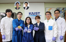
< From left: Professor Jongse Park , M.S candidate Jaehong Cho, M.S candidate Hyunmin Choi, Professor Brandon Reagen ISPASS >
Operating Large Language Model (LLM) services like ChatGPT requires a server infrastructure on the scale of tens of thousands of units. However, constructing actual equipment every time a new AI semiconductor or system architecture needs to be verified incurs massive costs and time. A research team at our university has developed a ‘virtual testbed’ that can pre-verify performance and efficiency inside a computer before building an actual large-scale AI server.
KAIST announced on May 29th that the research on a Large Language Model (LLM) serving infrastructure simulator (virtual testing software) developed by Professor Jongse Park’s research team in the School of Computing won the Best Paper Award at ‘ISPASS 2026 (IEEE International Symposium on Performance Analysis of Systems and Software),’ a world-renowned conference in the field of computer system performance analysis.
‘LLMServingSim 2.0,’ developed by the research team, is a simulation platform capable of virtually analyzing various hardware and software combinations in complex AI service environments. Researchers and developers can freely experiment with various design options and verify performance without having to directly build expensive, large-scale server infrastructures.
< LLMServingSim 2.0 is workload >
In particular, this technology is drawing attention because it goes beyond the existing Graphics Processing Unit (GPU)-centric environment to support diverse hardware environments, including Neural Processing Units (NPUs), which are rising as next-generation AI semiconductors, and Processing-In-Memory (PIM, a semiconductor technology that performs operations inside the memory).
In other words, it is a technology that allows future-oriented AI semiconductors that have not yet been commercialized to be tested in advance within a virtual datacenter environment. Through this, it is possible to replicate and analyze inside a computer how much the service speed improves, how much power consumption is reduced, and whether it operates stably even in a server environment scaled to tens of thousands of units when a specific semiconductor is applied.
In addition, it reproduces complex operations that occur during actual AI service operations—such as data processing, request distribution, and memory utilization—at the system level, enabling performance evaluations that are close to reality. Notably, it can even analyze disaggregated infrastructure environments where multiple server resources are separated and connected for use, showing great potential for utilization in next-generation AI datacenter research.
This simulator is expected to be widely utilized not only by researchers but also by LLM service companies and AI semiconductor startups to design and optimize next-generation AI infrastructures. This is because it can rapidly verify new AI semiconductors or service architectures prior to actual construction, thereby significantly reducing the cost and time of AI infrastructure development.
< Research Image (AI-generated image) >
Professor Jongse Park said, “The competitiveness of AI services is determined not only by the model itself but also by the infrastructure technology that operates it stably and efficiently.” He added, “We hope this simulator will serve as an important foundation for researchers and the industry to develop next-generation AI infrastructures faster and more efficiently.”
This research was led by M.S candidate Jaehong Cho and Hyunmin Choi in the School of Computing as co-first authors. Following their Best Paper Award at the 2024 IISWC (IEEE International Symposium on Workload Characterization), the research team won the Best Paper Award again at this ISPASS 2026, proving their research competitiveness in the field of AI infrastructure once more.
※ Paper Title: LLMServingSim 2.0: A Unified Simulator for Heterogeneous and Disaggregated LLM Serving Infrastructure, DOI: 10.1109/ISPASS69572.2026.00012 (Authors: Jaehong Cho, Hyunmin Choi, Guseul Heo, Jongse Park) ※ Open Source Link: https://llmservingsim.ai/ Meanwhile, this research was conducted with support from the Ministry of Science and ICT (MSIT), the Institute for Information & Communications Technology Planning & Evaluation (IITP, No. RS-2024-00396013), the Electronics and Telecommunications Research Institute (ETRI, No. RS-2025-02305453), and SK hynix.
KAIST Develops AI Technology That Automatically Ge..
<(From Left) Hyun-Bin Oh, Takida Yuhta, Uesaka Toshimitsu, Tae-Hyun Oh, Mitsufuji Yuki>
When people watch a scene in the film Jurassic Park where a giant dinosaur walks toward them, they naturally imagine a heavy, rumbling sound, as if the ground were shaking. This is because humans predict sound by considering not only the shape of an object, but also physical properties such as its size, weight, and speed of movement. However, existing video-to-audio generation AI mainly generates sound based on the category of objects or scene information in the video, and has not sufficiently reflected physical properties that vary depending on weight or speed.
KAIST (President Kwang Hyung Lee) announced on the 26th of May that a collaborative research team involving Professor Tae-Hyun Oh of the School of Computing, KAIST, together with joint researchers from POSTECH (President Sung Keun Kim) and Sony AI, has developed “PAVAS (Physics-Aware Video-to-Audio Synthesis),” an artificial intelligence (AI) technology that understands the physical situation in a video and generates more realistic sound.
<Concept Diagram of PAVAS (Physics-Aware Video-to-Audio Synthesis) Technology>
The key feature of this technology is that it is designed so that AI can infer invisible physical information such as the mass and velocity of objects in a video on its own. Ordinary videos do not provide exact numerical values for an object’s weight or speed, but the research team enabled AI to estimate them by analyzing the surrounding environment and movement context, and to reflect the results in the sound generation process.
In other words, the AI was designed to go beyond simply recognizing “what is visible” and to understand the physical cause of “why this sound should occur.”
As a result of technical validation, the research team’s AI generated sounds very similar to real-world environments in scenes involving physical interactions such as collisions or impacts between objects. In particular, it produced more realistic audio in which loudness and tone naturally changed when the mass and velocity of objects varied.
Recently, generative AI technologies that simultaneously generate video and audio have been advancing rapidly. Representative examples include Google’s “Veo 3” and ByteDance’s “Seedance 2.0.” However, in actual film, advertising, and game production sites, there is far greater demand for post-production work that adds sound effects suited to existing video scenes or supplements audio than for generating entirely new videos.
While existing commercial AI models have focused on generating video and audio together, PAVAS is differentiated by its ability to analyze the movement and collision characteristics of objects in a video and generate realistic sound effects that precisely match the scene.
<Comparison of Spectrograms Generated by Conventional Video-to-Audio Models and PAVAS>
The research team explained that this technology presents new possibilities in the field of “Physical AI,” or physically consistent generative AI. Physically consistent generative AI refers to AI that goes beyond simply producing plausible results and understands the laws of physics and causal relationships in the real world.
In the future, this technology is expected to provide more immersive user experiences in a wide range of fields, including the automation of content sound production, augmented reality (AR) and virtual reality (VR) content, the metaverse, and robotics simulation.
Professor Tae-Hyun Oh stated, “While existing generative AI has developed by increasing the scale of data and models, this research is meaningful in that it was designed so that AI directly understands physical quantities and causal relationships,” adding, “In the future, it can be expanded into a core foundational technology for next-generation multimodal AI that simultaneously understands and processes diverse types of information, including text, video, and speech.”
This study was led by POSTECH integrated M.S.-Ph.D. student Hyun-Bin Oh as the first author, with KAIST Professor Tae-Hyun Oh and Sony AI researchers Yuhta Takida, Toshimitsu Uesaka, and Yuki Mitsufuji participating as co-authors. This research was selected as an Oral presentation paper at CVPR 2026 (Computer Vision and Pattern Recognition 2026), the world’s most prestigious academic conference in the field of computer vision (image-based artificial intelligence technology), where only the top 0.88% of all papers are selected for oral presentation, recognizing the excellence of the work. The presentation is scheduled to take place on June 6.
※ Paper title: “PAVAS: Physics-Aware Video-to-Audio Synthesis,” DOI: https://arxiv.org/abs/2512.08282
This research was supported by the Mid-Career Research Program under the Basic Research Program of the Ministry of Science and ICT, the Pioneer Research Program for Future Converging Technology of the Ministry of Science, ICT and Future Planning, the AGI Program of the Ministry of Science and ICT, and the KAIST InnoCORE Program.
Talking to AI Before Seeing a Doctor… KAIST Develo..
<(Front row, from right)Professor Uichin Lee, Professor Eunjoo Kim, Professor Tak Yeon Lee, (Back row, from left) M.S candidate Gyeongmin Na, Ph.D candidate Yugyeong Jung, Researcher Hyangkyeong Oh, M.S candidate Jae Young Choi, Ph.D candidate Hyun Seung Moon>
People often say that seeking psychiatric care can feel intimidating. Patients may feel burdened when they first open up about their emotional distress, while medical staff must accurately understand a patient’s extensive history and symptoms within limited consultation time. Korean researchers have developed artificial intelligence (AI) technology that supports the initial psychiatric interview process, the first step in psychiatric care.
KAIST (President Kwang Hyung Lee) announced on the 24th of May that a joint research team led by Professor Uichin Lee of the School of Computing and Professor Tak Yeon Lee of the Department of Industrial Design, together with Professor Eunjoo Kim’s team from the Department of Psychiatry at Gangnam Severance Hospital (President Yong-Wook Kim), has developed a large language model (LLM)-based technology to support initial psychiatric interviews.
This study was conducted in a way that allows patients to first talk with AI before meeting a doctor, helping them organize their symptoms and condition in advance.
<AI Interviewer System Overview Diagram>
<Ask-Evaluate-Check-Plan Conversation Flow>
The research team designed the system so that AI can adjust the flow of conversation according to patient responses. The AI analyzes patients’ answers in real time by comparing them with specialized medical knowledge in psychiatry and generates the key questions that should be asked next. In particular, this system goes beyond simple question-and-answer interaction by applying real counseling techniques such as expressions of empathy, restating the patient’s words in an organized way, and clarifying ambiguous content. This is intended to help patients talk about their condition more comfortably.
As a result of experiments conducted with 1,440 virtual patients to verify performance, the team confirmed that in most cases, the system effectively obtained key clinical information needed for treatment within just 30 minutes.
Based on the collected conversation, the AI generates a clinical dashboard that shows symptoms and potential conditions at a glance and provides it to medical staff. Through this, doctors can understand the patient’s condition more systematically before the patient enters the consultation room, allowing them to focus more on in-depth counseling with the patient during the actual consultation.
The core of this research is that AI is defined not as a replacement for doctors, but as a “coachable apprentice ” It is a collaborative model in which AI handles repetitive and structured information collection, while doctors make the final diagnosis and prescription based on that information.
The research team made clear that AI still has limitations in understanding subtle emotional changes or handling sensitive topics, and emphasized that final judgment must always be carried out by trained medical professionals.
Professor Uichin Lee stated, “If AI reduces the burden of the initial consultation stage, medical staff can focus more on deeper counseling with patients,” adding, “This shows the possibility of developing a new model of care in which humans and AI collaborate in medical settings.”
This study, with doctoral student Yugyeong Jung as the first author, was presented on April 13 at ACM CHI 2026 (ACM Conference on Human Factors in Computing Systems), the most prestigious conference in the field of human-computer interaction.
※ Paper title: “Toward Flexible Psychiatric History-Taking and Visualization: Exploring Clinician Perspectives with Large Language Models,” DOI: https://dl.acm.org/doi/10.1145/3772318.3790970
※ Author information: Yugyeong Jung (KAIST, first author), Thu Hoang Anh Vo (KAIST, second author), Hyun Seung Moon (KAIST, third author), Jae Young Choi (KAIST, fourth author), Hyangkyeong Oh (Gangnam Severance Hospital, fifth author), Ujin Lee (Gangnam Severance Hospital, sixth author), Eunjoo Kim (Gangnam Severance Hospital, seventh author), Tak Yeon Lee (KAIST, corresponding author), Uichin Lee (KAIST, corresponding author)
This research was supported by the Digital Columbus Project of the Institute of Information & Communications Technology Planning & Evaluation (project title: Development of Digital Innovation Element Technologies for Predicting Complex Diseases in Advance and Expanding Non-Face-to-Face Care).
Overcoming the Limits of Hydrogen Storage and Tran..
<(Top row, from left) Professor Kang Taek Lee, Professor Joongmyeon Bae, Dr. Tae Ho Shin, Dr. Ki-Min Roh, (Bottom row, from left) Dr. Dongyeon Kim, Researcher Dong Jae Park, Dr. Incheol Jeong>
As ammonia gains attention as a next-generation energy source capable of overcoming the limits of hydrogen storage and transport, KAIST and a joint research team have developed fuel cell technology that directly uses ammonia as fuel while achieving world-class performance and stability. This achievement is regarded as a core technology that will accelerate the commercialization of the next-generation hydrogen economy and carbon-free power generation.
KAIST (President Kwang Hyung Lee) announced on the 20th of May that Professor Kang Taek Lee and Professor Joongmyeon Bae of the Department of Mechanical Engineering, together with a joint research team including Dr. Tae Ho Shin of the Korea Institute of Ceramic Engineering and Technology (KICET, President Jong-Suk Yoon) and Dr. Ki-Min Roh of the Korea Institute of Geoscience and Mineral Resources (KIGAM, President Kwon Yi Kyun), have developed catalyst technology that dramatically improves the performance and durability of ammonia-based protonic ceramic fuel cells (PCFCs, next-generation high-efficiency fuel cells that generate electricity by transporting hydrogen ions).
<AI image: A next-generation fuel cell that generates electricity using ammonia (NH₃) as fuel>
Ammonia is attracting attention as a next-generation hydrogen carrier (Energy Carrier, a medium that stores and transports hydrogen) because it is easy to store and transport in liquid form. It is also regarded as a representative carbon-free fuel because it consists only of nitrogen (N) and hydrogen (H), producing almost no carbon dioxide (CO₂) during power generation. However, inside fuel cells, ammonia has caused problems by damaging nickel-based materials and slowing reaction rates, leading to performance degradation and shortened lifespan.
To solve this problem, the research team designed a new catalyst structure combining a “high-entropy” oxide catalyst (High-Entropy, a design method that enhances material stability and performance by mixing multiple elements) that improves structural stability by mixing multiple elements, with metal nanoparticles (Nano Particle, ultrafine metal particles on the nanometer scale) that form spontaneously on the surface during operation.
This catalyst was found not only to resist structural collapse even in an ammonia environment, but also to effectively promote the reaction that decomposes ammonia into hydrogen. Through density functional theory (DFT, Density Functional Theory, a simulation method that calculates reaction mechanisms at the atomic level) analysis, the research team identified that the high-entropy oxide structure lowers the energy barrier required for ammonia decomposition and promotes the formation of metal particles.
<AI-generated image of a high-entropy catalyst made by mixing multiple metallic elements>
In particular, the metal alloy nanoparticles that formed spontaneously on the catalyst surface showed much higher catalytic activity than single-metal catalysts. A fuel cell applying this catalyst recorded a maximum power density of 2.04 W per unit area (1 cm²) at 700°C. This means that high power can be produced from an area the size of a fingernail, representing world-class performance in the field of ammonia-based protonic ceramic fuel cells that generate electricity by transporting hydrogen ions (protons).
In addition, the cell operated stably for more than 255 hours even under harsh conditions of 600°C, significantly improving the problem of performance degradation (a phenomenon in which performance decreases over time) seen in existing catalysts.
<Schematic of an ammonia-fueled PCFC incorporating a high-entropy catalyst>
<Microstructure and elemental distribution results of the high-entropy catalyst>
Professor Kang Taek Lee stated, “Through the synergistic structure of high-entropy oxides and alloy nanoparticles, we improved both the performance and durability of ammonia fuel cells,” adding, “This study will serve as a catalyst for accelerating the commercialization of ammonia-based carbon-free power generation technology and next-generation hydrogen energy systems.”
This research, with Dr. Dongyeon Kim of the Department of Mechanical Engineering at KAIST, researcher Dong Jae Park of the Korea Institute of Ceramic Engineering and Technology, and Dr. Incheol Jeong of the Korea Institute of Geoscience and Mineral Resources as co-first authors, was published on April 17 in Nano-Micro Letters (IF: 36.3), an international journal in the fields of energy and materials.
※ Paper title: “Entropy-Modulated Oxide–Metal Catalyst Architectures for Direct Ammonia Protonic Ceramic Fuel Cells,” DOI: https://link.springer.com/article/10.1007/s40820-026-02194-9
This research was supported by the Mid-Career Researcher Program of the Ministry of Science and ICT, the Global Basic Research Laboratory Program, the InnoCORE Program of the Institutes of Science and Technology, and the Basic Research Project of the Korea Institute of Geoscience and Mineral Resources.
<(From Left) Hyun Bae Bang, Cheon Woo Moon, Cindy Pricilia Surya Prabowo, Minjung Ki, Sang Yup Lee, Changhee Cho, (Upper Left) Jae Sung Cho, Namjin Jang>
KAIST announced on May 19th that the KAIST-Hanwha Solutions Future Technology Research Institute, has secured bio-technology capable of mass-producing eco-friendly raw materials for plastics and textiles using waste resources, offering an alternative to petroleum-derived naphtha.
Naphtha, an essential feedstock for the petrochemical industry, has faced sharp price increases and supply instability in recent years, driving demand for sustainable alternatives. The new technology, addresses both resource supply stability and environmental concerns simultaneously.
A study led by Distinguished Professor Sang-yup Lee of the Department of Chemical and Biomolecular Engineering was published on May 12th in the journal Nature Chemical Engineering and has been selected as the cover paper for the May issue, a designation reserved for research achievements that represent the corresponding issue.
This platform uses ‘glycerol,’ a byproduct discarded during the biodiesel production process, as a raw material. The team engineered high-efficiency microorganisms to convert this waste into 1,3-propanediol (1,3-PDO), a key material for plastics and cosmetics, and optimized the fermentation process for industrial application. The research team succeeded in maintaining high production level even in a 300L pilot process, which serves as a test production stage before application in large-scale plant facilities, moving beyond the laboratory scale.
This study also used computer simulations to predict which genes to engineer, which resulted in improved production levels. The team also developed the fermentation system without antibiotic supplementation — a significant advance, as antibiotic use in industrial fermentation raises concerns about antimicrobial resistance and regulatory hurdles for food, cosmetic, and pharmaceutical applications.
< (AI Image) Microbial-based process for 1,3-propanediol (1,3-PDO) production >
The achievement reflects a 10-year partnership between KAIST and Hanwha Solutions that began in November 2015, with researchers from both sides working together directly on the experiments. Through the KAIST-Hanwha Solutions Future Technology Research Institute, the collaboration has produced 6 patent applications and 13 published papers, standing as a representative model of industry-academic cooperation in South Korea.
< Schematic diagram of microbial-based metabolic engineering strategies for 1,3-PDO production >
※ Paper Title: High-titer, antibiotic-free, pilot-scale production of 1,3-propanediol by engineered Corynebacterium, DOI: 10.1038/s44286-026-00389-w
※ Authors: Jae Sung Cho (KAIST, First Author), Cindy Pricilia Surya Prabowo (KAIST, First Author), Taehee Han (KAIST), Cheon Woo Moon (KAIST), Yoo-Sung Ko (KAIST), Changhee Cho (Hanwha Solutions), Je Woong Kim (KAIST), Won Jun Kim (Hanwha Solutions), Hyun Bae Bang (Hanwha Solutions), Jae Eun Lee (KAIST), Minjung Ki (KAIST), Namjin Jang (Hanwha Solutions), Sang Yup Lee (KAIST, Corresponding Author)
Jung-dae Kim, Head of the Research Institute at Hanwha Solutions, said, “This research is highly significant in that it confirmed the possibility of replacing existing petrochemical processes using bio-based raw materials. We expect it to be an important foundation for sustainable chemical material production and industrial application in the future.”
KAIST Distinguished Professor Sang Yup Lee of the Department of Chemical and Biomolecular Engineering stated, “This research is a case showing that microorganism-based chemical production can be sufficiently expanded to an actual industrial scale beyond the laboratory. It will contribute to producing various chemical materials in a more eco-friendly way in the future.”
KAIST Wins Best Paper Award at Top HCI Conference ..
<(From Left) Professor Chang Hee Lee, Ph.D candidate Yoonji Lee>
A new type of digital game has emerged in which plants themselves change characters in the game, while humans observe and emotionally engage with them.
KAIST announced on the 15th of May that a research team led by Professor Chang Hee Lee of the Department of Industrial Design won the Best Paper Award at ACM CHI 2026, the most prestigious conference in the field of Human-Computer Interaction (HCI), for research that uses plants not as simple decorations or sensors but as “agents of interaction.”
< Plant.play system image >
< Plant.play system side view photo >
ACM (Association for Computing Machinery) CHI (Conference on Human Factors in Computing Systems) 2026 was held from April 13 to 17 in Barcelona, Spain. CHI is one of the world’s most prestigious international conferences in the field of Human-Computer Interaction (HCI).
The Best Paper Award is the highest honor awarded to only about the top 1% of all submitted papers. In particular, this year’s conference received a total of 6,730 paper submissions, marking the largest scale in its history, and this award is regarded as an achievement demonstrating the global research competitiveness of KAIST researchers.
Professor Chang Hee Lee’s team proposed a new form of interaction in which plants directly participate in digital games through the paper “When Plants Play: Rethinking Plant Materiality in Digital Games.”
This research is characterized by a design that goes beyond the conventional approach of using plants simply as sensors or decorative elements, allowing changes in the plant’s state to directly affect the progress of the game. The research team reflected the plant’s bioelectrical signals, environmental data, and circadian rhythms (biological changes that repeat according to day and night) in the game, enabling the character in the game to change according to the plant’s state. Rather than directly controlling the game, users participate by observing and interpreting the plant’s changes and responses.
As the plant grows, it creates different forms of characters and changes, and these changes reflect the plant’s own growth patterns and pace of transformation.
As a result of conducting user research in an actual exhibition environment, the research team confirmed that participants accepted the plant’s slow and unpredictable changes as a form of “play.” In particular, participants also showed a tendency to become emotionally immersed in and empathize with both the plant and the virtual character in the game. This led them to perceive the plant not simply as an object of observation, but as an entity with which they interact.
<A digital pet raised by the plant shows various behaviors—such as reading books—according to its daily rhythm, and grows over time>
<In a low-humidity environment, the plant provides hamburgers to the digital pet to help it grow>
This research received high recognition for moving beyond human-centered digital interaction and proposing new possibilities for interaction with nonhuman entities such as plants.
Professor Chang Hee Lee stated, “This research is an attempt to view nonhuman entities such as plants as agents and explore new forms of interaction,” adding, “Our society is expanding into an ‘attachment economy,’ which values emotional bonds and empathy, and in the future, emotional engagement not only with humans but also with diverse nonhuman entities such as AI, robots, animals, and plants will become important.” He continued, “This research is an example that demonstrates these new possibilities for interaction.”
This study, with doctoral student Yoonji Lee as the first author and Professor Chang Hee Lee as the corresponding author, can be found in the ACM Digital Library.
※ Paper title: “When Plants Play: Rethinking Plant Materiality in Digital Games”
※ DOI: https://doi.org/10.1145/3772318.3791373
This research was supported by Brain Korea (BK21).
<(From Left) Professor Inkyu Park, Dr. Seokjoo Cho, (Upper Right, From Left) Professor Ji-Hwan Ha, Researcher Junho Jeong , Professor Wei Gao>
“Diabetic ulcers,” which occur in patients with diabetes, are dangerous complications that can lead to amputation if the treatment window is missed. A joint research team has developed a “smart dressing patch” that can monitor wound conditions in real time.
KAIST (President Kwang Hyung Lee) announced on the 14th of May that a research team led by Distinguished Professor Inkyu Park of the Department of Mechanical Engineering, through joint research with Professor Ji-Hwan Ha of Hanbat National University (President Yongjun Oh), researcher Junho Jeong of the Korea Institute of Machinery & Materials (President Seog-Hyeon Ryu), and Professor Wei Gao of the California Institute of Technology (Caltech; President Thomas F. Rosenbaum) in the United States, has developed a “wireless, battery-free optoelectronic multi-modal sensor patch” for diabetic ulcer management.
The patch developed by the research team combines an optoelectronic sensor, which can simultaneously measure multiple types of biological information, with a functional dressing. It can analyze glucose concentration, acidity (pH, an indicator of hydrogen ion concentration), and temperature changes at the wound site in real time, and patients can check their condition themselves using a smartphone.
The research team fabricated a functional nanofiber dressing using electrospinning, a method that uses an electric field to create fibers much thinner than a human hair. This dressing changes color in response to increased glucose and changes in acidity that appear in diabetic foot wounds. In other words, if the wound condition worsens, the dressing color changes, allowing danger signals to be easily checked with the naked eye. Through this, abnormal signs that could lead to tissue necrosis can be detected and tracked over long periods in a non-invasive manner, meaning without cutting the skin or drawing blood.
The research team combined this with an optoelectronic system to improve diagnostic accuracy. A light-emitting diode (LED, a semiconductor device that converts electricity into light) embedded in the patch and a photodiode, a semiconductor sensor that detects light, measure the color change of the dressing as light reflectance and then convert it into an electrical signal.
This provides more accurate and stable data than ordinary camera-based imaging because it is less affected by changes in surrounding lighting.
In particular, the patch operates without a separate battery by applying a flexible circuit based on near field communication (NFC), a wireless communication technology that exchanges data over short distances. When a smartphone is placed near the sensor, the patch receives power wirelessly and operates, transmitting the measured data in real time. In other words, patients and medical staff can immediately check and respond to wound conditions using only a smartphone app, without separate complex equipment.
< Conceptual Diagram of a Multimodal Colorimetric Dressing and Optoelectronic Sensor for Diagnosing Diabetic Foot and Diabetic Diseases >
The technology developed in this study has high clinical value because it provides both intuitive visual signals and quantitative electronic data while imposing no physical burden on patients. It is also expected to contribute to improving the quality of life of patients with diabetes by enabling continuous wound management without repeated blood sampling.
Distinguished Professor Inkyu Park stated, “Research that began to reduce the pain of diabetic patients who have to prick their fingers with a needle every day has led to a technology for the preemptive diagnosis of complications,” adding, “This technology will become a core platform technology that can be expanded in the future to blood-free diagnostic technologies not only for diabetes but also for various chronic diseases.”
In this study, KAIST Dr. Seokjoo Cho and Professor Ji-Hwan Ha of Hanbat National University participated as co-first authors. The research results were published on March 26, 2026, in the international materials science journal Advanced Functional Materials. The paper was also selected as a Front Cover article of the journal.
※ Paper title: “Wireless, Battery-Free, Optoelectronic, Multi-Modal Sensor Integrated With Colorimetric Dressing for Diabetic Ulcer Management,” DOI: 10.1002/adfm.202532167
< Front Cover Image >
This research was supported by the Mid-Career Researcher Program of the National Research Foundation of Korea (NRF) funded by the Ministry of Science and ICT, the Alchemist Project of the Ministry of Trade, Industry and Energy, and the Daejeon RISE Center.
“Even Recognizing Puddles at Night”... KAIST Surpa..
< (From left) Ph.D. student Hanbin Cho, Postdoctoral Researcher Wenxuan Zhu, Professor Joonki Suh, and MS-PhD integrated student Changhwan Kim >
A technology that surpasses the limitations of existing sensors, which failed to distinguish between water and asphalt on dark roads, has emerged to enhance the accuracy of autonomous driving and medical diagnostics. Our university's research team has developed a next-generation polarization sensor that can read the "direction" of light and change its own response. KAIST announced on May 12th that a research team led by Professor Joonki Suh from the Department of Chemical and Biomolecular Engineering has developed a "self-reconfigurable" polarization sensor array technology that regulates its operation by finding the optimal state using "polarization" information—the property of light vibrating in a specific direction. With the recent explosive increase in data and the rapid development of artificial intelligence technology, the need for next-generation vision systems that can efficiently process vast amounts of information with low energy is growing. However, existing image sensors only detect the intensity (brightness) of light, limiting their ability to precisely grasp the orientation or surface structure of objects. To overcome these limitations, the research team developed a polarization-based sensor technology capable of recognizing the vibration direction of light. In particular, by utilizing a "heterostructure" that combines two different materials—tellurium (Te) and rhenium disulfide (ReS₂)—they effectively implemented characteristics where the response to light varies depending on the crystal orientation.
< Conceptual diagram of self-reconfigurable polarization sensor and in-sensor computing based on dual-anisotropy vdW heterostructures >
To precisely stack the two materials so they cross each other, the research team applied "Epitaxial Atomic Layer Deposition," a process that controls crystal structures by stacking materials precisely at the atomic layer level. By ensuring the crystal structures of the two materials interlock accurately, they secured higher reproducibility and stable performance compared to previous methods. In this structure, when light is irradiated, interfacial carrier transfer and trapping (a phenomenon where electrons move or stay at specific locations) occur at the material boundary. As a result, a "bipolar photoresponse"—a light-induced reaction where the current direction flips depending on conditions such as light intensity, wavelength, and direction—appears. A key feature is that the sensor's operating state can be freely adjusted using only light, without external electrical signals. Furthermore, this technology can be applied to "in-sensor computing" structures where the sensor itself processes data, allowing for the efficient processing of multi-dimensional optical information that changes over time without complex calculation processes. In actual experiments, it recorded a high accuracy of over 95% in recognizing moving objects, proving its potential for applications in various fields such as autonomous driving and medical diagnosis.
< Experimental image of a polarization AI sensor platform capable of light-based operational reconfiguration (AI-generated image) >
Professor Joonki Suh stated, "This research presents a new foundation for AI vision technology that can secure richer visual information by utilizing polarization information. It is expected to play an important role in implementing low-power, high-efficiency AI systems in the future." Wenxuan Zhu (Postdoctoral Researcher) and Changhwan Kim (Ph.D. student) participated as first authors in this study, with Professor Joonki Suh participating as the corresponding author. The research results were published on April 14 in the international academic journal Nature Sensors.
Paper Title: Self-reconfigurable polarization perception in dual-anisotropy heterostructures for high-dimensional in-sensor computing
Authors: Wenxuan Zhu, Changhwan Kim, Ruofan Zhang, Mingchun Lu, Namwook Hur, Hanbin Cho, Jihyun Kim, Jiacheng Sun, Joohoon Kang, Junchi Yan, Yuan Cheng & Joonki Suh
DOI: https://doi.org/10.1038/s44460-026-00057-9
< Paper portfolio and QR code >
Meanwhile, this research was conducted with the support of the PIM AI Semiconductor Core Technology Development (Device) Project and the Individual Basic Research Project of the National Research Foundation of Korea, funded by the Ministry of Science and ICT, and the Industrial Innovation Talent Growth Support Project of the Korea Institute for Advancement of Technology (KIAT).
KAIST demonstrates ultralow-noise microwave and mi..
<(From Left) Prof. Jungwon Kim, Dr. Changmin Ahn>
Researchers at KAIST have demonstrated a chip-scale photonic approach for generating ultralow-noise and highly stable microwave and millimeter-wave signals based on optical frequency combs (microcombs), offering a potential pathway toward compact, high-performance frequency sources for next-generation technologies.
High-frequency signals in the tens to hundreds of gigahertz range are essential for emerging applications such as 6G communications, radar, and precision sensing. However, achieving both low noise and high stability at these frequencies remains a fundamental challenge for conventional electronic signal sources.
In the first study, the researchers addressed the long-standing challenge of transferring the stability of an optical reference to a microcomb. Direct stabilization is difficult due to the lack of carrier-envelope offset detection in high-repetition-rate microcombs. To overcome this, they used a mode-locked laser as a transfer oscillator and synchronized it to the microcomb using electro-optic sampling. This approach enabled direct and robust transfer of optical-reference stability to the microcomb repetition rate, achieving fractional frequency stability at the 10-18 level and a phase noise of -125 dBc/Hz at 100 Hz offset from a 22 GHz carrier, representing state-of-the-art performance and more than 80 dB improvement over the free-running microcomb in the low-offset-frequency regime.
In the second study, the team addressed the degradation of noise performance typically observed when scaling microcombs to higher repetition rates. While microcombs with lower repetition rates (large resonators) exhibit better noise characteristics, increasing the repetition rate generally leads to performance degradation. The researchers showed that this limitation can be overcome using perfect soliton crystal (PSC) states, which enable repetition-rate multiplication while preserving the low-noise characteristics of the original comb. As a result, they generated millimeter-wave signals at 44 GHz and 66 GHz with timing jitter on the order of 3 femtoseconds, demonstrating that the low-noise performance of a microwave-rate microcomb can be preserved during scaling to millimeter-wave frequencies.
<Ultra-compact optical resonator chip with noise suppression based on an optical reference signal and increased frequency via fully solitonic waves (AI-generated image)>
Together, these results establish two key capabilities: (1) high-fidelity transfer of optical-reference stability to chip-scale microcombs, and (2) preservation of low-noise performance during frequency scaling to millimeter-wave regimes. This combined capability provides a practical route toward compact photonic signal sources that integrate optical-level stability with high-frequency operation.
The research was led by Dr. Changmin Ahn and Prof. Jungwon Kim at KAIST, in collaboration with Prof. Hansuek Lee. The results were published in Laser & Photonics Reviews and Optica.
· Optical-to-microcomb stability transfer for ultrastable timing and microwave/millimeter-wave generation (DOI: 10.1002/lpor.71135)
· Preserving ultralow timing jitter in microcombs with repetition-rate multiplication via perfect soliton crystal formation (DOI: 10.1364/OPTICA.581054)
First Observation of the Moment Lithium Batteries ..
< (From left) Professor Seungbum Hong, Ph.D candidate Seonghyun Kim, Dr. Youngwoo Choi, and Dr. Yoonhan Cho >
A crucial clue to simultaneously increasing electric vehicle (EV) driving range and battery lifespan has been discovered. A research team at our university has observed the exact moment of degradation in lithium metal batteries at the nanoscale (approximately 1/100,000th the thickness of a human hair) and identified the fundamental cause of performance decline. This is evaluated as a significant turning point in accelerating the commercialization of next-generation batteries.
KAIST announced on May 10th that a research team led by Professor Seungbum Hong from the Department of Materials Science and Engineering has identified the degradation mechanism of the lithium metal anode, a core component of next-generation batteries.
Lithium metal is dubbed a "dream battery material" due to its significantly higher energy density compared to conventional batteries. However, the rapid decline in performance after repeated charge and discharge cycles has been the biggest obstacle to commercialization. In particular, when lithium is deposited or stripped irregularly, it can form "dead lithium"—lithium that is electrically disconnected—which leads to performance degradation and poses safety risks.
The research team utilized in situ electrochemical atomic force microscopy (EC-AFM), which allows for real-time observation of the battery interior, to track the entire process of lithium deposition (plating) and removal (stripping). As a result, they confirmed that the lithium reaction does not occur uniformly across the entire surface but occurs selectively at specific locations.
<Overview of the EC-AFM Measurement Process>
Specifically, in porous regions with rough surfaces, voids were easily formed when lithium was stripped away, leading to the creation of "dead lithium" that becomes electrically isolated. This phenomenon acts as a direct cause of the sudden decline in battery performance.
The significance of this study lies in experimentally identifying where and how lithium metal batteries are damaged. Furthermore, it proved that the "initial morphology," where lithium is first formed, is a key variable that determines the long-term lifespan of the battery.
<Height Maps and Surface Slope Maps During the 1st–3rd Plating/Stripping Processes>
Accordingly, it is expected that if the surface where lithium forms is controlled uniformly and precisely in the future, battery life and stability can be dramatically improved. This suggests a design direction that can simultaneously achieve increased EV driving range and the development of long-life batteries.
Professor Seungbum Hong stated, "This research is highly significant as it directly confirmed the cause of battery performance degradation at the nanoscale. It will serve as an important foundation for developing safer and longer-lasting next-generation batteries."
Seonghyun Kim, a PhD student in the Department of Materials Science and Engineering, participated as the lead author. The study was published on February 24, 2026, in ACS Energy Letters, a prestigious international academic journal in the fields of materials science, chemistry, and chemical engineering, and was selected as a cover article.
※ Paper Title: Spatially Selective Lithium Plating and Stripping in Lithium Metal Anodes, DOI: https://doi.org/10.1021/acsenergylett.6c00122
< Photo of Selection as ACS Energy Letters Cover Paper >
Meanwhile, this research was conducted with support from LG Energy Solution and the Future Pioneering Convergence Science and Technology Development Program (RS-2023-00247245) of the National Research Foundation of Korea, funded by the Ministry of Science and ICT.
<(From Left) Ph.D candidate Jeesoo Yoon, Dr. Jinwon Oh, Professor Hee-Tae Jung, Professor Matteo Cargnello>
A KAIST and Stanford University joint research team revealed research results that overturn long-standing beliefs in the field of nanomaterials. Contrary to the conventional perception that mixing more metals complicates the system, this study revealed for the first time that complex compositions actually create more uniform nanoparticles, signaling a new turning point for next-generation energy and catalysis technology.
KAIST (President Kwang Hyung Lee) announced that a joint research team led by distinguished professor Hee-Tae Jung from the Department of Chemical and Biomolecular Engineering and Professor Matteo Cargnello from Stanford University has identified a paradoxical phenomenon where mixing more metals leads to the formation of more uniform nanoparticles.
Nanoparticles are core materials in various industries such as semiconductors, eco-friendly energy, and biotechnology. However, as the number of constituent elements increases, the different reaction rates of each element cause variations in particle size and shape, which has been considered a major challenge for precision control.
The research team focused on composition-focusing, a phenomenon where the particle components converge in one direction and become more uniform as the number of metal elements increases.
<Figure 1. (Top) Increasing number of possible product metal combinations (Bottom) Composition-focusing behavior>
The research confirmed that during the competitive bonding process of different metal atoms, the atoms that settle first act as a stepping stone, helping subsequent atoms attach more easily. Consequently, instead of mixing randomly, the atoms stack orderly in layers to form a stable structure. This phenomenon is a significant discovery, showing that the complex chemical reaction environment – previously viewed as a hurdle – actually helps atoms achieve an organized structure.
<Figure 2. (Top) Formation mechanism of five-element nanoparticles, (Bottom) comparison of catalytic performance>
To verify this principle, the team produced a multimetallic nanoparticle catalyst containing five different metals. In the reaction of decomposing ammonia to produce hydrogen – which requires high temperatures and high-performance catalysts – the new material showed four-times higher efficiency than the ruthenium catalyst, the current industrial standard.
Distinguished professor Hee-Tae Jung stated: This research is significant in that it discovered an unexpected paradoxical phenomenon and identified its operating principle. By utilizing this principle, we can design metal compositions tailored to desired performance, which is expected to be used in developing high-performance catalysts and eco-friendly energy materials for processes like hydrogen production and carbon dioxide conversion.
<Figure 3. Schematic illustration of multicomponent nanoparticle formation>
Jeesoo Yoon, a PhD candidate at KAIST, and Dr. Jinwon Oh from Stanford University participated as co-first authors of this study. The research was led by Distinguished Professor Hee-Tae Jung of KAIST and Professor Matteo Cargnello of Stanford University as co-corresponding authors. BASF (Badische Anilin- & Soda-Fabrik) and Seoul National University also participated in the joint research. The findings were published in the world-renowned academic journal Science on May 7th.
※ Title: Competitive reactivity drives size- and composition-focusing in multimetallic nanocrystals
※ DOI: 10.1126/science.aea8044
This research was conducted with the support from the National Research Foundation, the Korea Institute of Energy Technology Evaluation and Planning, and BASF.
KAIST Solves Computer Problems That Would Take Tho..
<(From Left) Professor Yang-Kyu Choi, Ph.D. candidate Seong-Yun Yun, (Upper Right) Professor Sanghyeon Kim, Dr. Joon Pyo Kim>
In the era of big data and artificial intelligence, a new approach has emerged for solving combinatorial optimization problems, which involve finding the most efficient solution among many possible options and can otherwise take thousands of years to compute. A KAIST research team has developed computational hardware that can be implemented entirely using existing silicon processes, enabling deployment on existing fabrication lines without additional facilities. This is expected to enable faster and more accurate decision-making across various industries, including logistics, finance, and semiconductor design.
KAIST (President Kwang-Hyung Lee) announced on the 6th of May that a joint research team led by Professor Yang-Kyu Choi and Professor Sanghyeon Kim from the School of Electrical Engineering has implemented an oscillatory Ising machine (a specialized-purpose computer in which multiple oscillating elements interact to find optimal solutions)—a next-generation specialized optimization hardware—using only conventional silicon semiconductor processes.
The research team focused on oscillators that repeat electrical signals periodically. As multiple oscillators exchange signals and synchronize their rhythms, the system naturally reaches the most stable state, and in this process, it finds the optimal solution.
Conventional oscillatory Ising machines have limitations in solving complex problems because it is difficult to precisely control slight frequency differences among oscillators, and the connectivity between elements is limited.
<An aging machine using a silicon oscillator and coupler>
To overcome this, the research team introduced a new approach in which both the oscillators and the couplers are implemented using single silicon transistors, which are the fundamental switching elements of semiconductors.
Through this approach, they reduced frequency deviations among oscillators, enabling stable synchronization, and by using couplers, they implemented multi-level coupling, allowing more precise reflection of problem weights.
As a result, both the ability to represent complex optimization problems and the performance of solution search were significantly improved. Using this technology, the research team successfully solved the representative combinatorial optimization problem known as the Max-Cut problem, which involves dividing a network into two groups to maximize connections.
This problem can be directly applied to various industrial fields such as logistics route optimization, financial portfolio construction, and semiconductor circuit placement. A key advantage of this approach is that it uses the CMOS* process currently employed in the semiconductor industry without requiring special materials or non-standard processes. Therefore, the technology suitable for mass production and commercialization on existing semiconductor production lines without additional facility investment.
*CMOS (Complementary Metal-Oxide-Semiconductor): the most standard process technology in modern semiconductor manufacturing, characterized by very low power consumption and low heat generation, and used to produce chips that serve as the “brains” of almost all digital devices, including smartphones and computer CPUs
<(AI-generated image) Concept diagram of an AI-based silicon aging machine>
Professor Yang-Kyu Choi stated, “This research presents an oscillatory Ising machine hardware that secures both scalability and precision by implementing both oscillators and couplers with silicon devices,” adding, “It is expected to be applied to various industrial fields requiring large-scale combinatorial optimization, such as semiconductor design automation, communication network optimization, and resource allocation.” He further noted that, as transistor miniaturization approaches its physical limits and increasingly requires atomic-level control, our group has spent the past decade exploring whether the future of transistors should extend beyond scaling toward the discovery of new functions. Futurist Alvin Toffler famously divided the development of society into three stages, describing the modern transition into a knowledge-based society as the “Third Wave.” In a similar way, the history of transistor technology, which now spans more than 80 years, may also be viewed in three waves. In 1935, Oskar Heil proposed the concept of controlling semiconductor current with an electric field in a British patent. In 1946, William Shockley developed the first solid-state transistor, an achievement that later led to the Nobel Prize. In 1961, Dawon Kahng invented the modern metal–oxide–semiconductor field-effect transistor, or MOSFET, which remains the foundation of today’s mass-produced semiconductor devices. From this perspective, the first wave of transistor technology can be defined as the “switch,” and the second wave as the “amplifier.” Our laboratory proposes a newly identified third wave: the transistor as an “oscillator.” For decades, semiconductor progress has largely been driven by improving the switching and amplification performance of transistors through miniaturization. However, as device fabrication now demands atomic-scale precision, the physical limits of scaling are becoming increasingly apparent. Future transistors therefore require a fundamental paradigm shift—from further miniaturization toward the realization of new functions. The greatest technological significance of this work lies in demonstrating the oscillator as a third fundamental function of the transistor. As a proof of this concept, we experimentally realized a physical Ising machine operating at room temperature.
This research was led by KAIST Ph.D. candidate Seong-Yun Yun and Dr. Joon Pyo Kim as co-first authors, and was published in Science Advances, one of the world’s most prestigious scientific journals, on March 27.
※ Paper title: “Scalable Ising machine composed entirely of Si transistors,” DOI: 10.1126/sciadv.adz2384
This research was supported by the National Research Foundation of Korea through the Next-Generation Intelligent Semiconductor Technology Development Program, the National Semiconductor Research Laboratory Core Technology Development Program, and the PIM Artificial Intelligence Semiconductor Core Technology Development Program.